Original article: Procarioti ed eucarioti: quali sono le differenze?, by Federica Angius
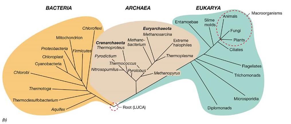
All living organisms can be divided into two groups, based on their cells’ structure: Prokaryotes and Eukaryotes. Prokaryotes include bacteria and archaea; small and simple ancient cells shape them. Eukaryotes involve algae, protozoa, protists, and fungus; arranged in more complex cells.
From prokaryotic cells to eukaryotic cells
National Institute of Health (NIH) says that prokaryotic organisms developed between 3.8 and 3.9 million years ago, while the eukaryotic organisms (animals and plants) have grown 2,7millions of years ago.
Nowadays, there are different theories about evolution. The working theory is called endosymbiotic theory, made at the end of the eighties by Linn Margulis, an American geneticist. According to this model, mitochondrion and chloroplasts came from ancient prokaryotes incorporated into more giant cells. This inclusion originated a symbiotic relationship, or rather a convenient collaboration, between two organisms that live one inside the other.
What do Prokaryotes and Eukaryotes have in common?
Although they are different, they share many components:
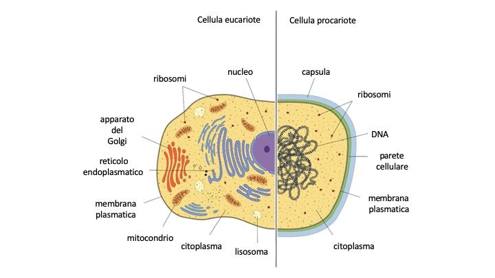
- Cytoplasmatic membrane: all the cells have a wall of permeability that divides the inside (cytoplasm) from the outside;
- Cytoplasm: an aqueous mixture of macromolecules like proteins, nucleic acids, and polysaccharides;
- Ribosomes: organelles involved in the synthesis of the proteins. Although the ribosomes in the eukaryotic cells are bigger, more complex, and bound by a membrane, in both the types of cells they are composed of two subunits: the big one the small one (respectively called the 60S and 40S in the Eukaryotes and 50S and 30S in the Prokaryotes);
- Cellular wall: based inside the cytoplasmatic membrane, sets up a firmer layer, giving structural strength to the cell. It is possible to find the cell wall in all the plant cells and many microorganisms while randomly in the animal cells.
What are the differences between Prokaryotes and Eukaryotes?
Nucleus/DNA
There are a lot of differences between Eukarya and Prokarya, but the bigger one is the nucleus. Eukaryotic cells have a double-wall delimited nucleus, interrupted by pores that let ions and macromolecules free to flow, allowing interactions between nucleus and cytoplasm. Inside the nucleus, there is the nucleoid. It is the most active part of the nucleus, where it is possible to find a considerable amount of enzymatic proteins and nucleic acids required to “read” the genetic code.
The prokaryotic cells do not possess the nucleus, but only a region called Nucleoid.
In the nucleus and nucleoid can be found all the biological history of the organism that it belongs to. DNA codes each information: like in Morse code, the succession of points and lines transmit a complete and complex message, so the sequences of chemical elements that form the DNA determine every cellular characteristic, and therefore, of the entire organism.
In the Eukarya, the DNA is divided into a couple of chromosomes, while in the Prokarya, the DNA forms the nucleoid that is circular and in a single copy. Moreover, most prokaryotes, besides their chromosome, have other circular DNA pieces called plasmids.
For example, Escherichia coli, the main prokatyotic organism, has 4288 genes, it is 7 times less then the genes content in the human cell (the human cell is eukaryotic).
Cellular organization
Organelles
The main characteristic of the eukaryotic cell is the presence of the organelles binded to the membrane:
- Mitochondrion: the “power stations” of the cell. Probably they come from bacteria that started to “live together” with the firsts nucleate cells from history. A double membrane delimits the mitochondrion that, bending inside, makes crests and tubules. On the inner membrane can be found the respiratory enzymes, while on the matrix that fills the internal/inward space, there are the substances necessary for the production of energy like sugars and fats;
- Lysosomes: spheroidal vesicles defined by a single membrane. They contain a high concentration of digestive enzymes that are used to destroy all the extra organelles or damaged or unusable molecules coming from the inside or the outside of the cell;
- Golgi apparatus: discovered by Camillo Golgi, an Italian scientist, in 1898. It is made of lipids, and it stores and revises proteins until the moment they are ready to be used or until they are expelled from the cell. The vesicles can flow in the membrane of other organelles and enrich them with their content, or they can merge with the plasmatic membrane and expel their material;
- Endoplasmic reticulum: thick net of double membranes, fold in different shapes that cross the entire cell. Located on these membranes, there is a complex of enzymes that allow specific biochemical reactions. Moreover, on its surfaces, the ribosome assembly and their presence give the typical “rough” appearance to this part of the reticulum. On the contrary, the part where there are few or none of them is called “smooth.” In this side of the reticulum, the production of the protein is absent or negligible;
- Hydrogenosomes: found in some anaerobic eukaryotic cells. Since these cells do not tolerate the O2, they replaced the mitochondrion with hydrogenosomes. They look like mitochondrion, but they do not have crests and a citric acid cycle. The organisms with the hydrogenosomes base their metabolisms on fermentation (like Trichomonas and the other protists). The primary biochemical reaction is the oxidation of the pyruvate in hydrogen (H2), carbon dioxide (CO2), and acetate;
- Chloroplasts: organelles that include chlorophyll that allows photosynthesis. It is possible to find them in eukaryotic phototrophic organisms. Like the mitochondrion, the chloroplasts have two membranes: the outer one, which is permeable, and the inner one that surrounds the internal matrix, called stroma, containing the principal enzymes for the Calvin cycle.
Cellular inclusions
Although the prokaryotes do not have organelles, often their cells possess inclusions visible at the microscope used to store energy, carbon or have special functions such as:
- Poly-b-hydroxybutyrate (PHB): thanks to the high quantity of carbon, it is synthesized by the cells (like in Ralstonia eutrophus or Bacillo megaterium). It is a lipid formed by units of poly-b-hydroxybutyrate that, after the polymerization, aggregate to form granules;
- Glycogen: a polymer formed by units of glucose, stock of carbon. Its energy is similar to PHB;
- Minerals of polyphosphate, sulfur, and carbonate: spare grains constituted by phosphate ions can be used to produce ATP. A lot of Gram-negative prokaryotes, like sulfur bacteria, can reduce the sulfur compounds. These bacteria accumulate sulfur that can be oxidized in sulfate when needed. Similarly, some cyanobacteria form carbonate minerals both in their outer and inner surface (like Gleomargarita). This process is called biomineralization;
- Magnetosome: intracell particles that form magretite chains. It gives a magnetic dipole to the cell, allowing them two orientations: the passive one and in a magnetic field. The magnetosomes make the magnetotaxis, a process in which the bacteria (that have magnetosomes) can dispose of themselves on the earth’s magnetic field lines.
Cytoskeleton
The eukaryotic cell is sustained by a thick frame formed by a complex net of fibrous proteins, with a strand shape that crosses the entire cell, giving its specific aspect and structure. Its function is to guide the movements of the organelles, so for this reason, it is essential in cellular division. The cytoskeleton net consists of microtubules, microfilaments, and intermediate filaments.
Cellular division
Eukaryotic cellular division has two processes: mitosis and meiosis.
Mitosis can be subdivided into different parts: condensation, divisions, and chromosomes’ separation in two groups, each one split into two daughter cells. It is typical in the yeast, for example, Saccharomyces cerevisiae (brewer’s yeast).
Meiosis is the conversion process from a diploid cell (two sets of chromosomes) to the haploid one (one set of chromosomes). It consists of two cellular divisions. In the first meiotic division, the homologous chromosomes segregate in two daughter cells, changing their genetic stage from diploid to haploid. The second meiotic division is very similar to mitosis, in which the haploid cells divided, forming a total of four daughter’s haploid cells. Prokaryotes divided for binary fission ( binary because from one cell become two). The prokaryotic chromosome replicates, and it is bound in two different parts of the membrane cell. The septum forces the cell to divide into two daughter cells, so, after its formation, the chromosomes separate. Basically, after the cellular division (cytokinesis), two identical cells are formed.
Bibliography
- Madigan, Martinko, Bender, Buckley & Stahl, Brock Biology of Microorganisms, 14th edition, Pearson.
- Rigutti A., Atlante di Fisiologia Umana. Il nostro corpo in azione dalle molecole agli apparati più complessi, seconda edizione, Giunti (link).